Brain scans now tie long COVID’s fatigue and brain fog to a measurable drop in dopamine signaling.
Story Snapshot
- Positron emission tomography scans found lower dopamine-related markers in long COVID patients.
- Reductions averaged about 16% to 20% across key motivation and movement hubs in the brain.
- Lower signals tracked with apathy, slowed movement, and memory issues reported by patients.
- The marker reflects dopamine nerve terminal integrity, often used in Parkinson’s research.
The Study That Put Numbers On Brain Fog
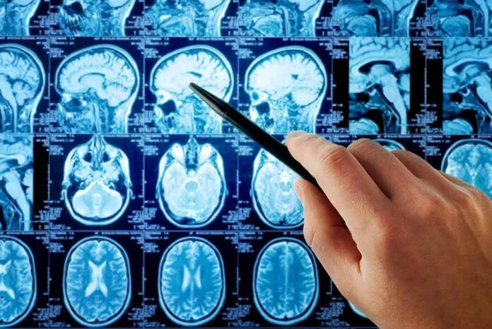
Researchers scanned 24 people with long COVID and 24 healthy peers. They used positron emission tomography to measure a protein called vesicular monoamine transporter 2, which marks dopamine nerve terminals. The long COVID group showed less binding in three striatal regions that govern motivation, movement, and memory. The differences were not small. Reported averages ranged from 16% to 20% lower than healthy controls, depending on the subregion measured.
The ventral striatum, which helps drive initiative and reward, showed the largest drop. The dorsal putamen and dorsal caudate also showed reduced signals. These areas help the brain plan actions and hold working memory online. The imaging readout does not guess at symptoms. It measures a system that many patients describe as “out of gas.” That tight link between signal and function is why this result landed with force across outlets.
Why Dopamine Terminals Matter In Long COVID
Dopamine tunes how we move, think, and pursue goals. Vesicular monoamine transporter 2 loads dopamine into tiny packets at nerve endings. When positron emission tomography shows less vesicular monoamine transporter 2, it points to fewer active terminals or a drop in terminal function. Nuclear medicine has used this marker for decades to index dopamine neuron integrity in diseases like Parkinson’s. That track record gives this signal real weight beyond a vague “brain change”.
Long COVID patients often report apathy, slowed movement, and memory slips. The scan patterns lined up with these complaints. Lower ventral striatum binding tracked with more apathy. Lower putamen binding tracked with slower movements. Lower caudate binding tracked with worse memory scores. That symptom-to-circuit match is the detail that turns a lab finding into a likely clinical clue rather than a trivia fact about proteins.
How Big Is A 16%–20% Drop?
A change of that size will not paralyze someone. But it can blunt the mental spark that gets you off the couch and keeps your thoughts clear under stress. Dopamine systems run like an amplifier. Less terminal capacity means less punch with the same input. That can feel like effort costs more and joy pays less. For a person who was fine before COVID and now drags through the day, that gap maps well to lived experience reported in clinics and surveys.
Researchers have found evidence that long COVID may damage dopamine releasing neurons in the brain, potentially explaining persistent fatigue, low motivation, slowed movement, and memory difficulties. The discovery could lead to new treatments that target https://t.co/YTeLtXkt5q
— Michael W. Deem (@Michael_W_Deem) September 22, 2026
Patients in the study had mild or moderate initial infections. That matters. The scan differences did not require a severe hospital stay or a ventilator. Many who now struggle with brain fog were never hospitalized. This research suggests a brain basis that matches their symptoms and their history. It also narrows the search for treatments toward dopamine circuits and the forces that disrupt them after infection.
What This Does—and Does Not—Prove
The key win here is biological footing. The study shows a reduced dopamine terminal marker tied to hallmark symptoms. That directs doctors and scientists to a specific circuit instead of a catch-all bucket. The result does not prove that dopamine neurons died. Vesicular monoamine transporter 2 positron emission tomography is an indirect readout. Lower binding can reflect fewer terminals, lower protein expression, or altered vesicle content. Still, as a screening light on the dashboard, it is bright and pointed.
Reports describe a single-center sample of 24 long COVID participants against 24 controls. That is tight but typical for positron emission tomography. The authors focused on people with clear neuropsychiatric symptoms, which likely boosted the signal. That framing means the result best fits that subtype, not every long COVID case. Even so, the alignment across outlets on the numbers and regions suggests the backbone finding is sound as reported.
What Comes Next For Patients And Clinicians
Clinicians can start where the data point. Screen for apathy, bradykinesia-like slowing, and working memory strain. Consider therapies that support attention, structured activity, and dopaminergic tone. Avoid overpromising a cure. If inflammation or immune triggers drive the change, fixing the circuit may also require calming the cause. Research should test whether vesicular monoamine transporter 2 signals recover as patients improve, and whether anti-inflammatory or pro-dopamine strategies shift the scan and the symptoms together.
Policy leaders should note the practical message. Long COVID is not only fatigue and mood. It includes a brain circuit problem that robs productivity and purpose. Support trials that compare subtypes, use the same tracer, and track patients over time. Tie imaging to blood markers to find who benefits from what. That path respects common sense: measure what is broken, treat to target, and prove it moved the needle in daily life.
Sources:
fortune.com, medicalxpress.com, gigazine.net, medscape.com